Skin Checks:


TESTING THESE COLOURS,,,,
Skin Cancer is the most common cancer and the incidence is increasing each year. Factors that indicate increased risk for melanoma skin cancer include overexposure to the sun, episodes of sunburn in childhood, personal or family history of skin cancer, having fair skin that burns easily and having a large number of moles. Regular checks are very effective in detecting skin cancers and allowing for treatment at an early stage.
Skin Clinic has the latest digital camera technology to ensure the most advanced technology available with "Skin Check". The aim of Skin Check is an early detection of Melanoma, as well as Basal Cell, Squamous Cell Skin Cancers and other rarer forms of skin cancer. Melanoma is the most critical as early detection of Malignant Melanoma can mean curative surgery for what is potentially a deadly disease.
The first step is body mapping the whole skin with the High Definition (HD) camera, giving us a baseline to work from and allowing the ability to review all areas of the skin for any future changes. Body mapping is an invaluable tool for the diagnosis of Melanoma as 70% of Melanoma arise as new moles. The TBI (Total Body Imaging) photos are copied to a CD to allow for self-checking between skin checks.
The second and most important step is to have one of our highly trained team conduct a full skin check. We are experts at spotting skin cancers when they are early and subtle, when others might overlook them. Obvious skin cancers are identified as well as moles that are 'atypical' and need to be monitored closely.
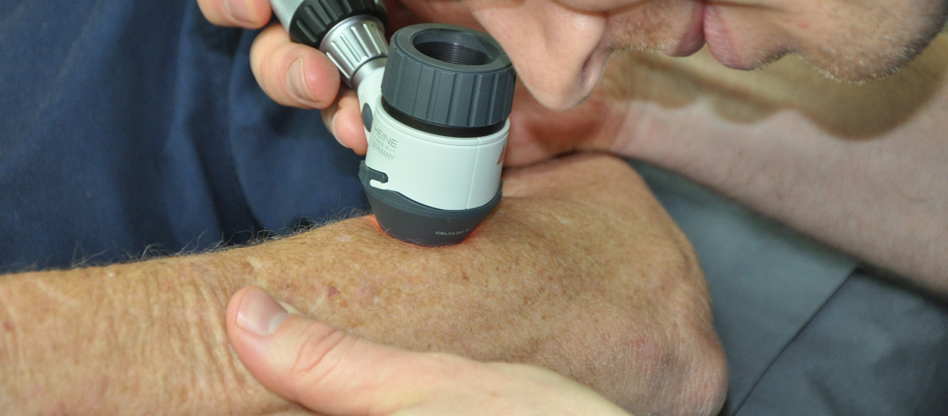
In the third step, lesions that are less dangerous but have a risk for evolving into cancer are then photographed through a dermatoscope with the HD camera. The dermatoscope allows us to see the exact structure of the mole. Dr Dowley and Dr Leslie, review all the highly magnified digitised images of the individual lesions, taking into account the clinical appearance and history.
A follow up appointment is made to repeat these close-up images at intervals appropriate to the nature of the mole. With two images side by side for comparison, subtle changes that can herald development of cancer within a lesion are able to be picked up.
Your skin is constantly changing so we recommend that you self-check every 3 months and have a professional check undertaken every year or two for peace of mind. Early detection makes the difference.